Fatty Liver Detection System
AI-powered classification of liver ultrasound images using Siamese Neural Networks (ResNet-50)
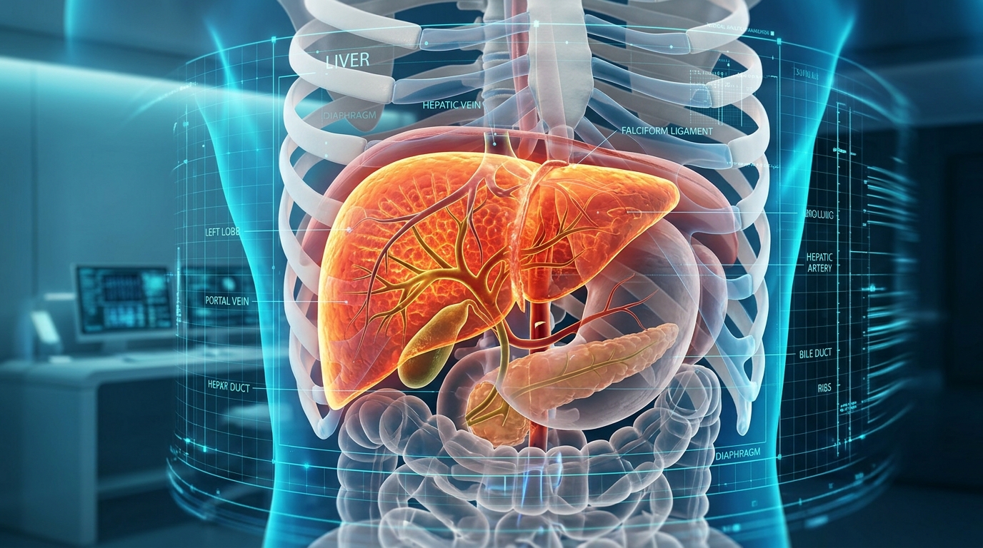
Image Classification
Classify liver ultrasound images into 5 categories
Deep Learning Model
Siamese Neural Network with ResNet-50 encoder
5-Class Prediction
Normal, Grade I, Grade II, Grade III, CLD
Fast & Easy to Use
Upload an image and get instant prediction results
Upload Ultrasound Image
Sample Ultrasound Image
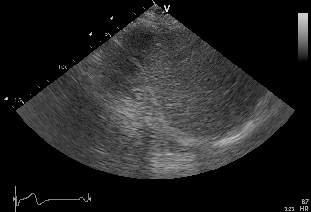
Predicted Class Probabilities
Ultrasound Analytics & Clinical Detail Graphs
Biomarker Engine ActiveQuantitative acoustic sound decay, multi-class probability metrics, parenchymal echogenicity, and steatosis radar
Prediction History
| Record ID | Date & Time | Filename | Mode | Prediction | Confidence | Severity | Action |
|---|
Siamese Neural Network Architecture
Twin ResNet-50 feature extractor with contrastive self-supervised pre-training and downstream 5-class linear classifier.
Supported Steatosis Categories
About the Fatty Liver Detection System
Non-Alcoholic Fatty Liver Disease (NAFLD) affects up to 25% of the global adult population and is strongly associated with metabolic syndrome, type 2 diabetes, and cardiovascular conditions. Early and accurate detection of hepatic steatosis is essential to halt or reverse progression toward non-alcoholic steatohepatitis (NASH), cirrhosis, or hepatocellular carcinoma.
While liver biopsy is the historic reference standard, it is invasive, costly, and associated with patient discomfort and complications. Ultrasound B-mode imaging is widely accessible and non-invasive, but visual qualitative evaluation suffers from inter-observer and intra-observer variability.
This clinical AI system implements a Siamese Neural Network with a ResNet-50 backbone trained via self-supervised contrastive learning. By extracting high-dimensional latent representations of liver parenchyma and ultrasound attenuation patterns, the model provides objective, reproducible assessments across all five clinical steatosis categories.
Clinical Usage Protocol
- Ensure the ultrasound image captures liver parenchyma adjacent to the right kidney or diaphragm.
- Verify the scan is in standard B-mode format (JPG, PNG, or BMP).
- Upload the scan or select one of the clinical reference samples.
- Review the multi-class probability distribution and confidence score.
- Export or print the report for medical records and radiologist corroboration.